Beyond the Mammogram: The Future of Breast Cancer Screening
Breast cancer screening is becoming more personal, with genetic testing, breast-density assessments and new imaging techniques helping doctors identify women who need closer monitoring. European research is already changing clinical practice, although national screening programmes have yet to agree on how widely to introduce the new methods.
A woman’s age still determines when most European health services invite her for breast cancer screening. Women typically enter national mammography programmes in their late forties or fifties, with examinations repeated at fixed intervals. Doctors arrange earlier surveillance for patients with certain inherited genetic mutations or a strong family history, although most women follow the standard schedule.
Research into breast density and inherited cancer risk has exposed weaknesses in that approach. A mammogram is less sensitive in extremely dense breast tissue, where small tumours are harder to distinguish from surrounding structures. Meanwhile, women without an obvious family history still account for most breast cancer diagnoses. Researchers are developing more precise ways to identify those who need closer monitoring, combining genetic information, breast-density measurements and individual medical histories.
European radiologists are already incorporating some of these findings into clinical practice. MRI screening has demonstrated benefits in women with extremely dense breasts, while trials in Sweden have shown that artificial intelligence improves cancer detection and reduces radiologists’ workloads. National health services must now decide how widely to introduce additional examinations without increasing unnecessary investigations.
What breast density reveals
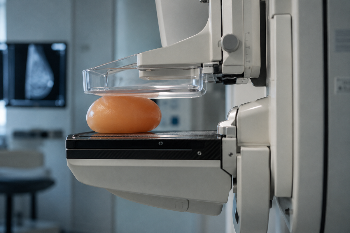
On a mammogram, fatty tissue appears dark, while glandular and fibrous tissue appear white. Tumours also tend to appear white, making abnormalities harder to identify when the surrounding breast tissue is dense. Radiologists classify density into four categories, ranging from almost entirely fatty breasts to extremely dense breasts. Neither breast size nor a physical examination establishes the classification.
Women with extremely dense breasts face approximately twice the breast cancer risk of women with average breast density, according to the European Society of Breast Imaging (EUSOBI). Their mammograms are also less sensitive, leaving a greater possibility that a cancer will remain undetected until symptoms develop or the next examination takes place.
Evidence from the Dutch DENSE trial prompted EUSOBI to recommend supplementary magnetic resonance imaging, or MRI, in 2022. The trial enrolled women aged 50–75 with extremely dense breasts whose mammograms had shown no suspicious findings. Researchers found that adding MRI detected cancers missed by mammography and reduced the number diagnosed between scheduled screening rounds.
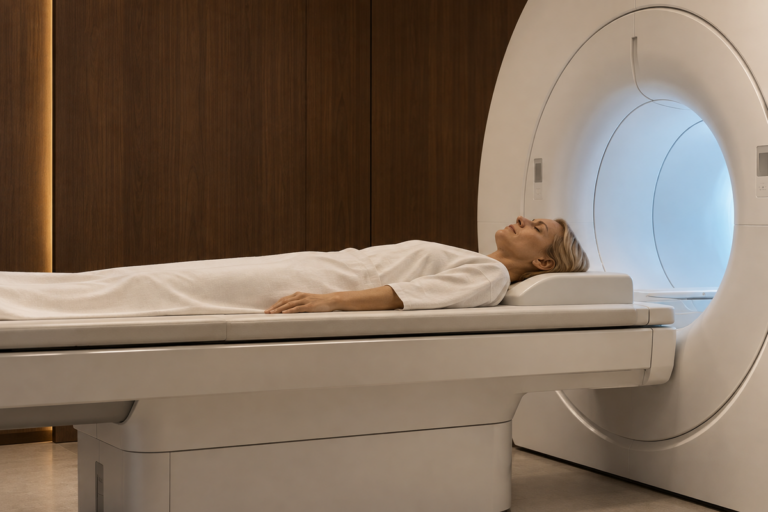
MRI uses magnetic fields and radio waves to produce detailed images of breast tissue, generally with an injected contrast agent that helps reveal suspicious areas. It is more sensitive than mammography, but the additional findings also bring more follow-up examinations and biopsies, including some that ultimately find no cancer.
EUSOBI recommends offering MRI screening every two to three years to women with extremely dense breasts. The European Commission has adopted a more cautious position on introducing supplementary MRI throughout organised screening programmes. Scanner availability, examination costs and the burden of additional investigations remain part of the assessment, alongside the clinical evidence.
Genetics and the women who need earlier screening
Some women enter specialist breast surveillance in their twenties after genetic testing identifies inherited mutations associated with a high risk of cancer. BRCA1 and BRCA2 are the best known, although variants in PALB2, CHEK2 and ATM also influence susceptibility. Their effects differ considerably, and doctors assess genetic findings alongside family history and other medical information.
Women carrying certain high-risk variants generally begin annual MRI examinations around the age of 25 or 30, depending on the mutation and national clinical guidance. Mammography is introduced later in some programmes. Genetic counselling accompanies testing because the results also have implications for relatives who share the same inherited variant.
Most breast cancers, however, occur without a recognised high-risk inherited mutation. Researchers have consequently developed statistical models that bring together a wider range of information. The Tyrer-Cuzick and BOADICEA models assess family relationships, reproductive history, previous breast conditions and, where available, breast density and genetic findings.
BOADICEA also incorporates polygenic risk scores, which estimate inherited susceptibility from numerous common genetic variations. Each variation generally has a small effect, but their combined influence helps distinguish between women whose estimated risks differ despite having similar family histories.
Doctors use these calculations to decide whether someone qualifies for earlier mammography, supplementary MRI or specialist assessment. A lifetime risk estimate of 20 percent, for instance, describes a probability rather than a diagnosis. The models also depend on accurate patient information and require validation across different populations. Researchers continue to examine how well genetic risk scores perform among women whose ancestry is underrepresented in the datasets used to develop them.
More than one way to examine the breast
Digital breast tomosynthesis, commonly known as 3D mammography, has already entered clinical practice in many countries. Instead of relying on conventional two-dimensional images alone, the technique produces a series of views from different angles. Radiologists examine thin sections of tissue to distinguish suspicious abnormalities from overlapping normal structures. Studies have found improved cancer detection and fewer recalls in some screening settings, although results vary between populations and screening programmes.
Another technique, contrast-enhanced mammography, uses an injected iodine-based agent to highlight areas with unusual blood supply. Doctors are studying its role in women who need further investigation but cannot undergo MRI because of medical contraindications, limited access or other practical difficulties. Its place in routine population screening remains less firmly established.
Ultrasound provides additional information when a mammogram reveals an abnormality or a patient reports a lump. It is also used to examine dense breast tissue, particularly where MRI is unavailable. The examination involves no ionising radiation, but supplementary ultrasound screening produces additional false-positive findings and does not match MRI’s sensitivity in high-risk screening.
Specialists select imaging according to the reason for the examination and the woman’s individual risk. Someone with a high-risk genetic mutation follows a different surveillance programme from a woman attending routine mammography, while an unexplained breast lump requires diagnostic investigation regardless of a recent normal screening result.
Artificial intelligence is changing the radiologist’s workload
Sweden’s MASAI trial has supplied some of the strongest clinical evidence for artificial intelligence in breast cancer screening. Approximately 106’000 women participated in the study, which compared AI-supported mammography with the conventional Swedish practice of having two radiologists independently assess each examination.
Researchers reported in 2025 that AI-supported screening detected 6.4 cancers per 1’000 women, compared with 5.0 per 1’000 in the standard screening group. Radiologists’ image-reading workload fell by approximately 44 percent, without a statistically significant increase in false-positive results.
An additional analysis published in The Lancet in January 2026 examined cancers diagnosed between screening examinations. AI-supported screening achieved a sensitivity of 80.5 percent, against 73.8 percent for conventional double reading, while specificity remained at 98.5 percent in both groups.
The trial met its prespecified statistical criterion showing that AI-supported screening was no worse than standard screening for interval cancer rates. Although researchers recorded fewer interval cancers in the AI group, the difference was not statistically significant.
Algorithms analyse mammogram images and identify findings that warrant closer assessment, allowing radiologists to concentrate attention on examinations where the probability of an abnormality is higher. The technology also helps departments manage growing screening volumes without increasing staffing at the same rate.
Hospitals nevertheless require evidence beyond initial detection figures. Researchers are continuing to assess the effects on advanced cancer diagnoses, overdiagnosis and mortality, while evaluating how AI systems perform across different imaging equipment and patient populations. An algorithm trained on one dataset does not automatically deliver the same performance in every screening programme.
Europe’s screening programmes face difficult choices
The European Union’s 2022 cancer-screening recommendation supports organised mammography for women aged 50–69, with countries given the option of extending programmes to women aged 45–49 and 70–74. European Commission guidance recommends screening every two years for average-risk women aged 50–69, while examining different intervals for younger and older age groups.
National health services retain responsibility for eligibility, funding and implementation. Their programmes differ in screening ages, examination intervals and access to specialist services, leaving women across Europe with different opportunities for early detection.
Introducing personalised screening across entire populations requires changes beyond the radiology department. Genetic counselling services need capacity to assess family histories and explain test results. Hospitals must arrange additional MRI examinations and follow-up appointments, while national information systems need to incorporate individual risk profiles without compromising patient confidentiality.
EUSOBI has encouraged breast cancer risk assessment from around the age of 25, principally to identify women whose family history or genetic circumstances warrant surveillance before routine mammography begins. The recommendation concerns evaluating risk and referring selected patients for specialist assessment, rather than offering mammograms or genetic tests indiscriminately to all young women.
Clinical trials are examining whether screening based on individual risk improves outcomes compared with fixed age-based schedules. The results will help determine how health services allocate additional imaging, how frequently lower-risk women receive examinations and whether more personalised programmes justify their expense.
MRI scanners are costly to operate and examinations take longer than mammograms. Greater sensitivity also brings additional findings that require investigation, some of which prove harmless. Health authorities must weigh those demands against the prospect of detecting more cancers at a stage when treatment is less intensive and survival prospects are better.
Prevention enters the discussion earlier
Women identified as having a high inherited or calculated breast cancer risk receive more than a revised examination schedule. Specialist teams also discuss measures intended to reduce the likelihood of developing the disease, including medication and, in selected circumstances, preventive surgery.
Tamoxifen and certain other medicines reduce the incidence of hormone-receptor-positive breast cancer among women at increased risk. Their adverse effects and contraindications require individual assessment, while risk-reducing surgery is generally reserved for women whose genetic or medical circumstances place them at particularly high risk.
Lifestyle remains part of prevention advice. Alcohol consumption is an established breast cancer risk factor, while regular physical activity and maintaining a healthy weight, especially after menopause, are associated with lower risk. These measures do not eliminate the possibility of developing cancer, and screening itself aims to detect disease rather than prevent it.
Women attending routine appointments already have access to information that helps doctors assess their circumstances. A mammogram establishes breast density, while a detailed family history, including cancer diagnoses on both sides of the family, identifies patterns that warrant closer attention. Previous medical treatment, particularly chest irradiation at a young age, also influences the assessment.
European hospitals now have stronger evidence for supplementary MRI in women with extremely dense breasts, more detailed genetic risk models and clinical trial results supporting AI-assisted mammography. Wider adoption depends on how national health services organise screening, fund specialist examinations and assess the consequences of detecting more abnormalities. The research has advanced considerably; translating it into consistent care across Europe remains unfinished.